The Role and Application of a Procto Sigmoidoscope in Modern Medicine
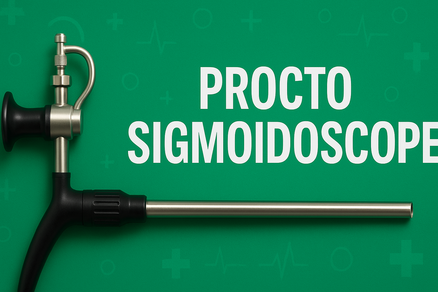
A Procto Sigmoidoscope often referred to as a rigid sigmoidoscope or proctosigmoidoscope is a medical instrument designed for direct visualization of the rectum and the lower part of the colon (sigmoid colon). At its most basic, it is a rigid tube equipped with lighting and optics; in more advanced modern forms, it integrates a camera and imaging system to provide video-assisted visualization. In practice, the procto sigmoidoscope is gently inserted via the anus, allowing physicians to visually inspect the inner lining of the rectum and distal colon, observe mucosal abnormalities, detect bleeding sources, take biopsies, or carry out minor therapeutic interventions. Its adoption and evolution reflect the convergence of traditional endoscopic techniques with digital imaging technology, enabling more precise diagnosis, better documentation, and enhanced patient care.
History and Evolution of Sigmoidoscopy
The concept of sigmoidoscopy dates back to the late 19th and early 20th centuries, when rigid instruments with simple optics were used to examine the lower bowel. Over decades, as fiber optics and flexible endoscopy developed, flexible sigmoidoscopes became more common. However, rigid scopes retained certain advantages in terms of durability, better image control, and ease of sterilization.
With advances in imaging sensors and miniaturization, modern rigid procto sigmoidoscopes now often include a “chip-on-tip” camera, integrated LED lighting, and digital signal pathways (e.g., USB output), merging the telescope, optics, light source, sheath, instrumentation channel, and video interface into a compact “5-in-1” device. These newer devices combine the sturdiness of rigid instruments with the advantages of real-time video and image capture—improving diagnostic yield, facilitating teaching or telemedicine, and enabling simplified documentation.
Anatomy of a Modern Rigid Video Procto Sigmoidoscope
A contemporary rigid video procto sigmoidoscope is constructed from medical-grade stainless steel and comprises several integrated components:
-
Tubular sheath / outer body: A hollow rigid tube, typically 30 cm in working length, through which the scope is inserted and maneuvered.
-
Obturator tip: A rounded, blunt tip to aid smooth insertion without trauma. In fixed-tip designs, the camera and lens are built into the tip itself.
-
Optical / camera system: Usually a miniature video camera placed at the distal end, coupled with LEDs (e.g. six LEDs) to illuminate the lumen.
-
Instrumentation channel: A side channel (or lumen) that allows passage of biopsy forceps, graspers, or other instruments.
-
Gas/air insufflation port: A side port with a Luer-lock connection for insufflating air or CO₂ to distend the lumen and improve visualization.
-
Digital interface / signal conversion: A circuit (often located centrally) that converts analog signals from the camera into digital output, commonly via a USB interface. This allows connection to computers, tablets, or display systems.
-
Handle / hub: A structural element that supports the camera electronics and provides the hub for instrumentation channels, wiring, and control.
Such devices may come in varying diameters—for example, 10 mm and 15 mm outer diameters—to suit patient variation and procedural needs. The tip usually provides zero-degree vision, meaning the optics look straight ahead. The device is reusable and designed for sterilization (e.g. by Ethylene Oxide or formalin exposure) between uses.
Indications: When and Why a Procto Sigmoidoscope is Used
A procto sigmoidoscope is used in a range of diagnostic and minor therapeutic settings, primarily relating to diseases of the lower colorectal tract. Key indications include:
-
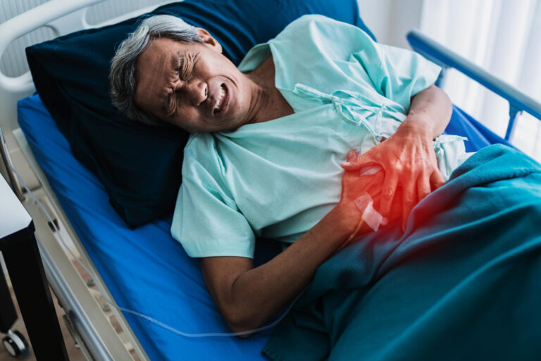
Investigation of rectal bleeding
When a patient presents with hematochezia (fresh blood in stool) or rectal bleeding, a proctosigmoidoscopy helps localize the source—be it hemorrhoids, ulceration, erosions, or neoplasia. -
Evaluation of anorectal symptoms
Persistent pain, tenesmus (feeling of incomplete evacuation), change in bowel habits, or mucus discharge may prompt a sigmoidoscopic evaluation to look for fissures, proctitis, strictures, or masses. -
Surveillance & screening
In patients with known colorectal pathology (polyps, prior cancer, inflammatory disease), periodic video proctosigmoidoscopy helps monitor for recurrence, new lesions, or dysplastic changes. -
Biopsy or sampling
The instrumentation channel allows passing biopsy forceps to take tissue samples from suspicious mucosa for histopathology. -
Minor therapeutic interventions
Some scopes allow limited therapeutic maneuvers: polypectomy, cauterization of bleeding points, band ligation (for hemorrhoids), or local coagulation of ulcers. -
Pediatric or special conditions
In certain congenital conditions (e.g. Hirschsprung’s disease), rigid scopes or video-enabled counterparts may be used to obtain biopsies or intervene in anorectal anomalies.
Procedure: How the Examination is Performed
A typical procto sigmoidoscopy using a rigid video scope proceeds as follows:
-
Patient preparation
The patient lies in the left lateral (or lithotomy) position, with knees drawn up. The digital rectal examination is first done to assess tone, tenderness, or palpable masses. -
Lubrication and insertion
The scope is lubricated and gently inserted. The rounded obturator helps reduce mucosal trauma. Insufflation (air or CO₂) is applied to expand the lumen and improve visibility. -
Advancement & inspection
The scope is slowly advanced to the sigmoid colon, observing the mucosa, vascular pattern, and any abnormal lesions. The integrated LED lighting provides illumination, while the video camera relays live feed to a display device connected via USB. -
Intervention / sampling
If a suspicious lesion is seen, biopsy forceps or other instruments are passed through the instrumentation channel to obtain tissue or even perform minor treatments. -
Withdrawal
The scope is slowly withdrawn, re‐inspecting the mucosa on exit. Any areas of interest are documented, and images or video segments may be saved. -
Post-procedure care
Patients may experience mild cramping or bloating (due to insufflated air). Light bleeding can occur, especially after biopsy. Most individuals can resume normal activity soon after, unless complications arise.
The procedure typically takes 5 to 15 minutes. It is tolerated well, with minor discomfort but seldom severe pain, though local anesthesia or sedation may sometimes be offered.
Advantages of Modern Video-Enabled Rigid Procto Sigmoidoscopes
Compared with older rigid devices or flexible instruments, modern video procto sigmoidoscopes offer several compelling benefits:
-
Integrated imaging and recording: The chip-on-tip or camera system enables high-quality, real-time visualization and allows still images or videos to be recorded, facilitating documentation, follow-up comparison, or remote consultation.
-
Compact and cost-effective: By combining the scope, optics, lighting, and instrumentation channel in one device, these instruments avoid the need for large, expensive separate video towers. Some models are marketed at a fraction of the cost of conventional video colonoscopy systems.
-
Robust and durable design: Rigid stainless-steel construction makes these instruments sturdy, easy to clean, and less prone to mechanical breakdown compared to flexible scopes.
-
Simplified setup and portability: With USB output, such scopes can connect to laptops, tablets or Android devices, enhancing portability and access in resource-limited settings or mobile clinics.
-
Improved sterility and reuse: The rigid design allows thorough sterilization between uses, preserving durability and reducing long-term maintenance costs.
-
Good procedural control: Rigid instruments provide tactile feedback and precision, which can be preferable in certain anorectal procedures compared to flexible scopes.
Limitations and Considerations
Despite their advantages, procto sigmoidoscopes also have limitations and must be used judiciously:
-
Limited reach: Rigid sigmoidoscopes cannot access the entire colon; they are confined to the rectum and lower sigmoid colon. More proximal lesions remain unseen.
-
Patient discomfort: Rigidity may cause discomfort, especially in patients with tight or spasmodic rectal muscles. Sedation or analgesia may be needed in sensitive individuals.
-
Risk of injury: Though designed with blunt tips, there is still potential for mucosal abrasion or perforation if forced or improperly handled.
-
Less flexibility: Unlike flexible scopes, rigid devices cannot navigate sharp bends or tortuous segments of the colon.
-
Dependency on operator skill: Proper interpretation, biopsy technique, and maneuvering require expertise. In inexperienced hands, the diagnostic yield may decline.
Clinical Applications: Real-World Cases
Let us consider some illustrative uses of procto sigmoidoscopy:
-
Case of rectal bleeding in a middle-aged patient
A 50-year-old patient presents with fresh blood on toilet paper intermittently for several weeks. A video proctosigmoscopy reveals an ulcerated polypoid lesion in the lower rectum. Biopsy is taken through the instrument channel; histopathology confirms adenocarcinoma. Prompt surgical referral is made. -
Surveillance in inflammatory bowel disease
In a patient with a history of ulcerative colitis limited to the rectum and sigmoid, periodic proctosigmoidoscopic checks allow monitoring for dysplasia or neoplastic transformation. -
Hemorrhoid intervention
Some scopes permit band ligation of internal hemorrhoids via the instrumentation channel, combining diagnostic and limited therapeutic function in a single sitting. -
Pediatric biopsy
In suspected Hirschsprung’s disease, mucosal or submucosal biopsies from the rectum can be obtained during the procedure for diagnostic confirmation.
Best Practices, Safety, and Recommendations
To maximize benefit and minimize risk, clinicians should observe several best practices:
-
Proper patient selection
Reserve rigid video procto sigmoidoscopy for cases where distal evaluation is sufficient and flexible colonoscopy is not needed (or is contraindicated). -
Adequate preparation
Bowel preparation (cleansing enemas) may be required to improve visualization. Adequate lubrication and slow insertion help reduce trauma. -
Gentle technique
Avoid force; insufflation should be moderate to avoid overdistention. Always advance and withdraw under direct vision. -
Sterilization protocols
The instrument must be sterilized per hospital protocols (e.g. Ethylene Oxide, formalin chamber) to prevent cross-infection. -
Training and competency
Physicians should undergo proper training in endoscopic anatomy, instrument handling, biopsy technique, and complication management. -
Documentation and archiving
Use the video capture features to archive images and videos for future reference, comparison, and consultation. -
Patient communication
Inform the patient about the procedure, possible sensations (cramps, bloating), and aftercare, including expectations of mild bleeding or gas discomfort.
The Future of Procto Sigmoidoscopy
As medical imaging and miniaturization evolve, procto sigmoidoscopes are likely to benefit from further improvements:
-
Higher-resolution imaging
Incorporation of HD, 4K sensors, or enhanced illumination (e.g. narrow-band imaging) may improve detection of subtle mucosal changes. -
Wireless / tetherless designs
Removing the need for wired USB connections could simplify workflow, though power and data transfer constraints would need addressing. -
Integrated AI / aided detection
Real-time image analysis and lesion detection algorithms might assist clinicians in recognizing polyps or subtle lesions during the procedure. -
Disposable or semi-disposable components
To further minimize infection risk and cleaning requirements, parts of the device (e.g. sheaths) might become single-use while maintaining core metallic components. -
Telemedicine and remote consultation
The video output could permit real-time consultation or oversight by remote specialists, enhancing care in underserved or rural areas. -
Enhanced working channels / miniaturized tools
New biopsy or ablation tools small enough to pass through slimmer instrumentation channels may expand the therapeutic potential.
Conclusion
The Procto Sigmoidoscope remains a vital tool in colorectal diagnostics and minor interventions, especially for lesions of the rectum and sigmoid colon. Modern rigid video procto-sigmoidoscopes bring together the robustness of traditional rigid scopes with the advantages of digital imaging, enabling efficient, documented, and patient-friendly examinations. While they do not replace full colonoscopy for complete colonic assessment, they play a crucial role in targeted evaluation, surveillance, and procedural simplicity. With ongoing technological advancements—higher image resolution, AI-aided detection, and wireless integration—the utility and accessibility of proctosigmoidoscopy are poised to grow further in clinical practice.