Understanding the Rigid Ureteroscope: A Complete Guide
The Rigid Ureteroscope is a specialized medical instrument that plays a central role in the diagnosis and treatment of urinary tract conditions. Used primarily by urologists, this tool has revolutionized how doctors manage issues within the ureter and parts of the urinary system. This article explores the design, function, clinical applications, advantages, limitations, and future developments related to the rigid ureteroscope, offering a comprehensive overview for both medical professionals and curious readers alike.
What Is a Rigid Ureteroscope?
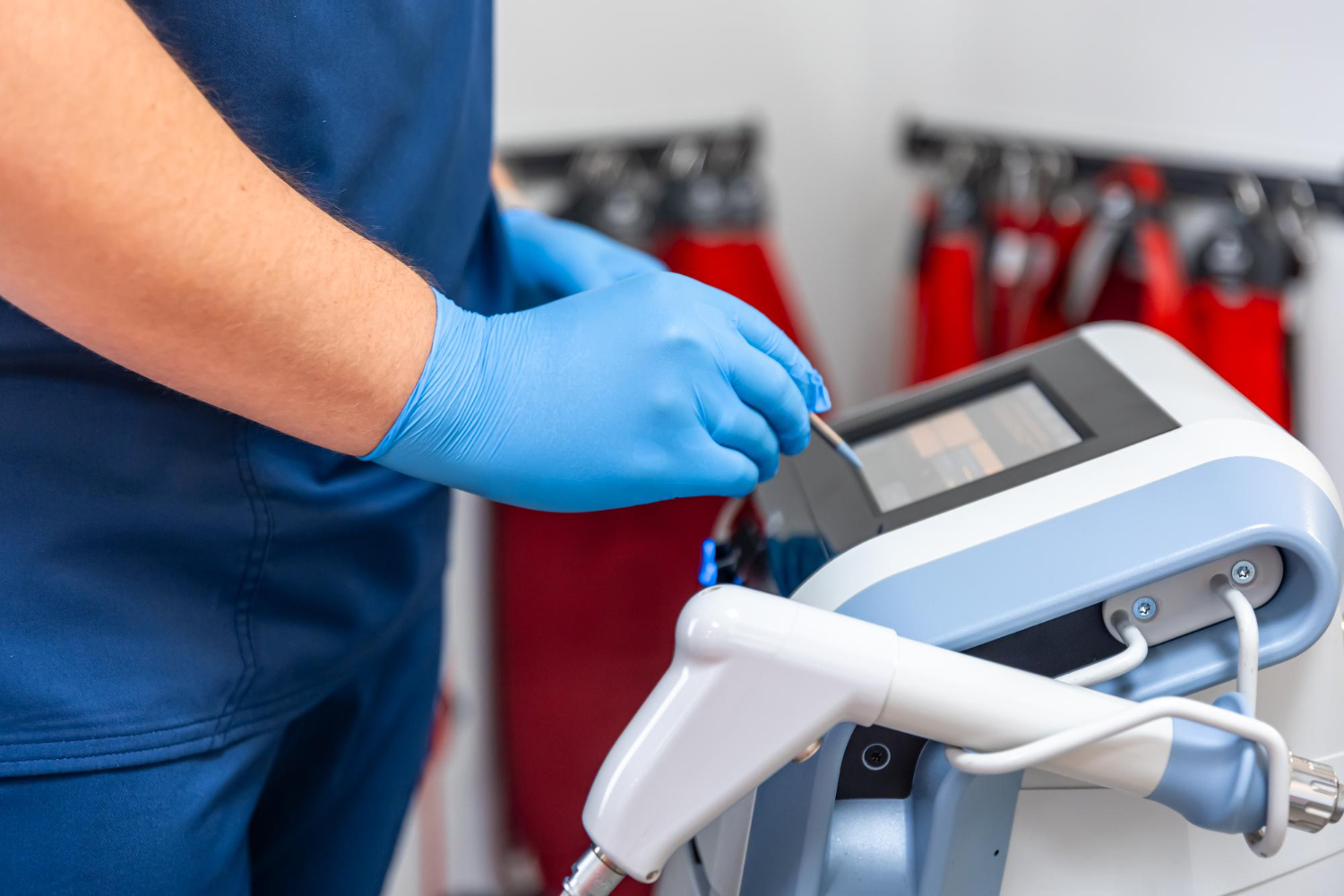
At its core, the rigid ureteroscope is a long, thin, straight endoscopic device designed to enter the urethra and pass through the bladder into the ureter—the tube that carries urine from the kidney to the bladder. Unlike flexible ureteroscopes, which can bend and navigate curved pathways, the rigid ureteroscope maintains a fixed straight shape. This structural characteristic directly affects how and when it is used in clinical practice.
The instrument consists of a metal or fiberoptic shaft, a viewing lens system, and a channel for passing surgical tools or irrigation fluid. The optical system allows the urologist to visualize the internal surfaces of the ureter directly. The ability to see the ureter’s interior in real time makes the rigid ureteroscope indispensable in diagnosing conditions such as strictures, stones, tumors, and other anomalies.
Historical Development
The rigid ureteroscope traces its origins to the broader field of endoscopy, which began in the 19th century. Early instruments were rudimentary and provided limited vision and functionality. Through advances in optics, lighting, and material science, modern rigid ureteroscopes now offer high-definition visualization and improved patient safety. Although flexible scopes have grown in popularity for many applications, rigid ureteroscopes remain widely used due to their durability and effectiveness in certain procedures.
Key Components and Design Features
A typical rigid ureteroscope includes the following essential parts:
-
Optical System: High-quality lenses and light transmission mechanisms provide clear images for accurate diagnosis and safe navigation.
-
Shaft: The straight, rigid metal or fiberoptic shaft allows precise control during insertion and manipulation.
-
Working Channel: A lumen through which instruments, such as baskets, laser fibers, or biopsy forceps, can be passed.
-
Irrigation Ports: These allow saline or other fluids to flush the area, improving visibility and assisting in stone removal.
The design also includes ergonomic handles and controls that help the clinician maneuver the scope and perform tasks such as stone retrieval or tissue sampling with precision.
Clinical Applications
One of the most significant uses of the rigid ureteroscope is in the management of ureteral stones. Ureteral lithiasis, or stones lodged within the ureter, can cause intense pain, bleeding, and obstruction of urine flow. The rigid ureteroscope allows direct access to these stones, enabling their fragmentation and extraction using auxiliary tools like laser fibers and retrieval baskets.
Beyond stone management, the rigid ureteroscope assists in diagnosing unexplained hematuria (blood in urine), locating strictures (narrowing of the ureter), and performing biopsies. In cases where a suspected tumor is present, the ability to visually inspect and sample tissue helps guide further treatment and management.
Another application of the rigid ureteroscope is in treating certain types of ureteral strictures. By visualizing the obstruction directly, the urologist can dilate the narrowed segment or perform surgical corrections under direct vision.
Procedure Overview
A typical procedure involving a rigid ureteroscope is performed under anesthesia. The patient is placed in a lithotomy position, and the scope is gently inserted through the urethra into the bladder. From there, it progresses into the ureter under direct visualization.
Once the target area is reached, the clinician may employ various tools through the working channel. For instance, stones can be fragmented using laser lithotripsy, after which fragments are removed. Biopsies can be taken, and stents may be placed if needed to ensure proper urine drainage post-procedure.
Throughout the procedure, continuous irrigation keeps the surgical field clear. When the work is complete, instruments are removed, and the scope is withdrawn carefully.
Advantages of Using a Rigid Ureteroscope
The rigid ureteroscope offers several notable advantages in urologic practice:
-
Superior Control: Its straight, sturdy design allows excellent tactile feedback and control during stone removal and manipulation.
-
High-Quality Visualization: Especially in proximal and mid-ureteral procedures, the optical clarity ensures accurate diagnosis and treatment.
-
Durability: Rigid instruments are less likely to sustain damage during use compared to more delicate flexible scopes.
-
Cost-Effectiveness: In many settings, the rigid ureteroscope can be more affordable to maintain and repair than flexible alternatives.
Because of these benefits, the rigid ureteroscope remains a go-to choice for many urologists handling stones and other conditions located in the lower and mid-ureter.
Limitations and Challenges
Despite its advantages, the rigid ureteroscope has inherent limitations. Its lack of flexibility means it cannot easily navigate curved or tortuous anatomy, especially in the upper ureter and renal pelvis. For patients with anatomical variations or in cases requiring access beyond a certain curvature, flexible ureteroscopes are preferred.
Another consideration is patient comfort and risk of injury. While generally safe, rigid scopes may cause more discomfort or ureteral trauma if not used with careful technique. Proper training and experience are crucial to minimize such risks.
Comparisons with Flexible Ureteroscopes
Flexible ureteroscopes have gained popularity because they allow easier navigation into difficult-to-reach areas within the urinary tract. These instruments can bend and adapt to curves, making them valuable for accessing the kidney’s intrarenal anatomy. However, flexible scopes are expensive, delicate, and require meticulous sterilization and maintenance.
In contrast, the rigid ureteroscope is rugged and better suited for straightforward ureteral work. Many urologists choose the rigid instrument for initial evaluation and intervention in straightforward stone cases, reserving flexible scopes for complex anatomy or upper-tract pathology.
Future Trends and Innovations
The field of ureteroscopy continues to evolve, with ongoing advancements aimed at improving visualization, maneuverability, and therapeutic outcomes. Emerging technologies may integrate high-definition imaging, enhanced optics, and digital platforms into rigid designs, merging some benefits of both rigid and flexible systems.
Robotics and augmented reality may also play roles in future procedures, offering greater precision and reducing surgeon fatigue. Miniaturization of components, improved ergonomic designs, and cost-effective manufacturing may further expand the accessibility of ureteroscopic techniques worldwide.
Training and Best Practices
Proper use of the rigid ureteroscope demands training and clinical experience. Urologists undergo specialized education in endourology, where they learn techniques for insertion, visualization, stone management, and complication avoidance. Simulation labs and continuing medical education help clinicians stay current with best practices and technological advances.
Preoperative planning and patient evaluation are critical. Imaging studies such as ultrasound, X-ray, or CT scans help determine stone size, location, and ureteral anatomy, guiding the choice of instrument and approach. Collaboration among surgical teams ensures patient safety and optimal outcomes.
Patient Considerations and Recovery
Patients undergoing procedures with a rigid ureteroscope typically experience quick recovery times, especially when minimally invasive techniques are used. Mild discomfort, burning during urination, or small amounts of blood in the urine are common temporary effects. Doctors often provide pain management, hydration advice, and follow-up instructions to support healing.
In cases where stents are placed, patients may experience irritation until the stent is removed. Regular follow-up appointments ensure that stones are fully cleared and that the urinary tract heals without complications.
Conclusion
The rigid ureteroscope remains a fundamental instrument in modern urology. Its durability, visual clarity, and precision in handling ureteral stones and other conditions make it an essential tool for many clinicians. While its design limits access to certain areas compared to flexible scopes, its advantages continue to uphold its value in clinical practice.
Understanding the role, application, and limitations of the rigid ureteroscope helps both medical practitioners and patients navigate decisions about urinary tract treatment options. As technology advances, we can expect further refinement of this foundational instrument—continuing to improve outcomes and expand the possibilities of endoscopic urinary care.